
Scleral Lens Optics: How Vault Design and Saline Physics Correct Your Vision
Scleral lenses correct vision by vaulting entirely over the cornea and filling the space between the lens and the eye with a saline reservoir that optically replaces the irregular corneal surface. Understanding how does optics of a scleral lens work helps you make a more informed decision about whether this technology fits your vision needs.
Key Takeaways
The saline reservoir between the lens and cornea acts as a fluid tear lens that neutralizes corneal irregularities before light enters the eye.
Scleral lenses rest on the sclera (the white of the eye), not the cornea, giving them exceptional stability and comfort.
Gas-permeable RGP material allows sufficient oxygen to reach the cornea despite the lens's large diameter.
Wavefront-guided scleral designs can correct higher-order aberrations that standard glasses and soft lenses cannot address.
Conditions like keratoconus, severe dry eye, and corneal scarring respond well to scleral lens correction.
Fitting scleral lenses requires multiple precise measurements and trial fittings to optimize vault, diameter, and optical zone.
What Scleral Lenses Are and Why Optometrists Prescribe Them
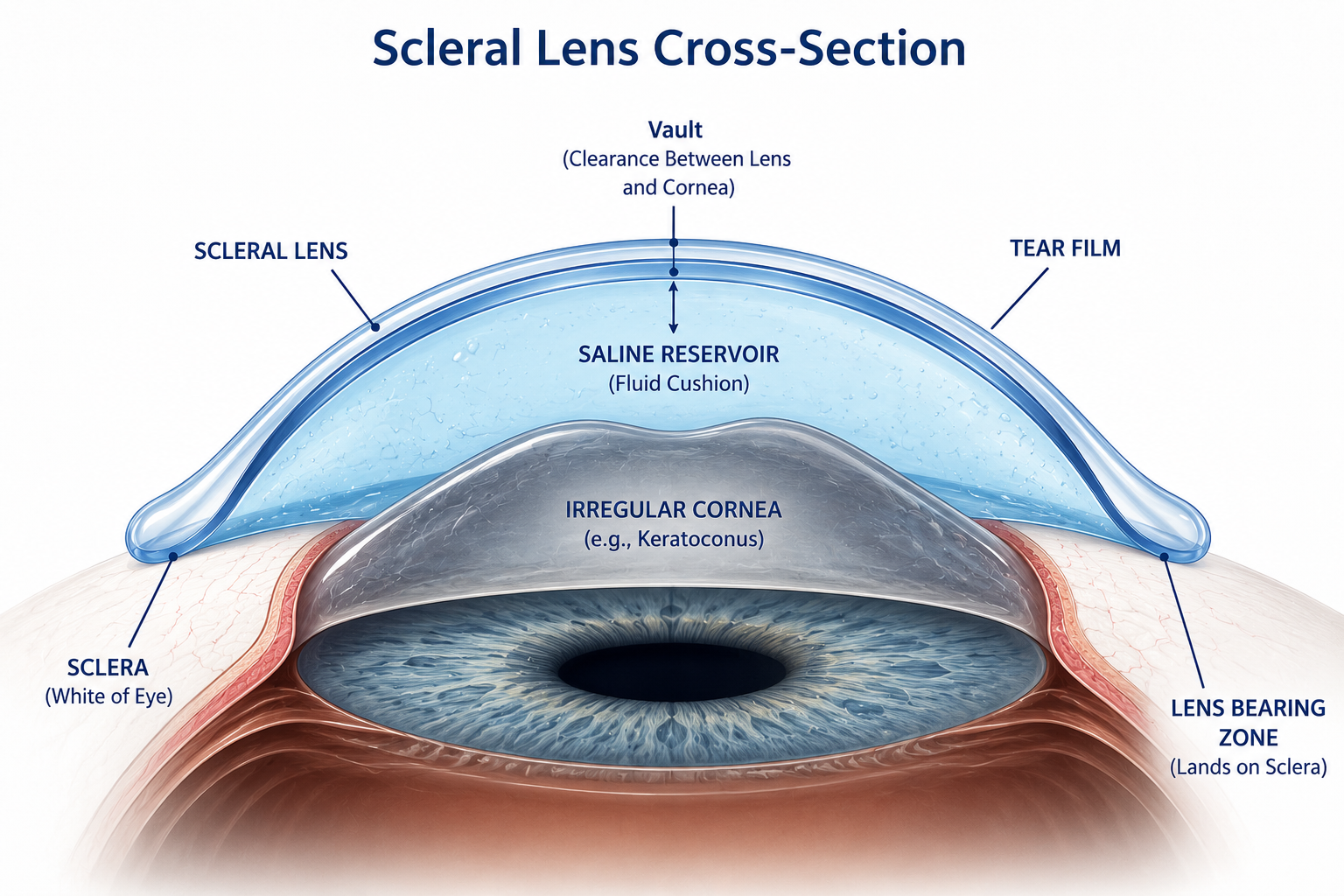
Scleral lenses are large-diameter, rigid gas-permeable (RGP) contact lenses, typically ranging from 14.5 mm to over 22 mm in diameter. Unlike standard soft lenses that drape over the cornea, scleral lenses arch entirely over it and land on the scleral tissue at the outer edge. That single design choice changes everything about how they interact with light and the ocular surface.
Optometrists prescribe them primarily for patients whose corneas are too irregular for conventional lenses to deliver functional vision. Conditions that commonly drive this decision include keratoconus, corneal ectasia after LASIK, pellucid marginal degeneration, corneal scarring, and severe refractive errors that glasses cannot adequately correct. They also serve patients with surface disease conditions such as Stevens-Johnson syndrome and chronic dry eye, because the fluid dome beneath the lens keeps the cornea continuously hydrated.
According to the American Academy of Ophthalmology, scleral lenses have become a leading non-surgical option for patients with keratoconus, a condition affecting roughly 1 in 2,000 Americans. You can explore what fitting these lenses looks like by reviewing the services offered at Texas Contact Lens Institute in Plano, TX.
The Core Optical Mechanics: Vault Geometry and the Saline Tear Lens
This is where how does optics of a scleral lens work becomes genuinely interesting from a physics standpoint. A standard cornea refracts light in a smooth, predictable arc. An irregular cornea, like one distorted by keratoconus, scatters light unpredictably, producing ghosting, halos, and distorted acuity.
The saline solution that fills the dome between the lens back surface and the corneal front surface has a refractive index very close to that of the corneal tissue itself. That similarity is critical. When light passes from the rigid lens surface into the saline layer and then into the cornea, the optical discontinuity at the corneal surface is essentially eliminated. The saline acts as a fluid optical element, smoothing over all the peaks and valleys of the irregular corneal topography before light ever reaches the corneal stroma. The result is that the front surface of the scleral lens becomes the primary refracting surface, and it is a perfectly smooth, engineered curve.
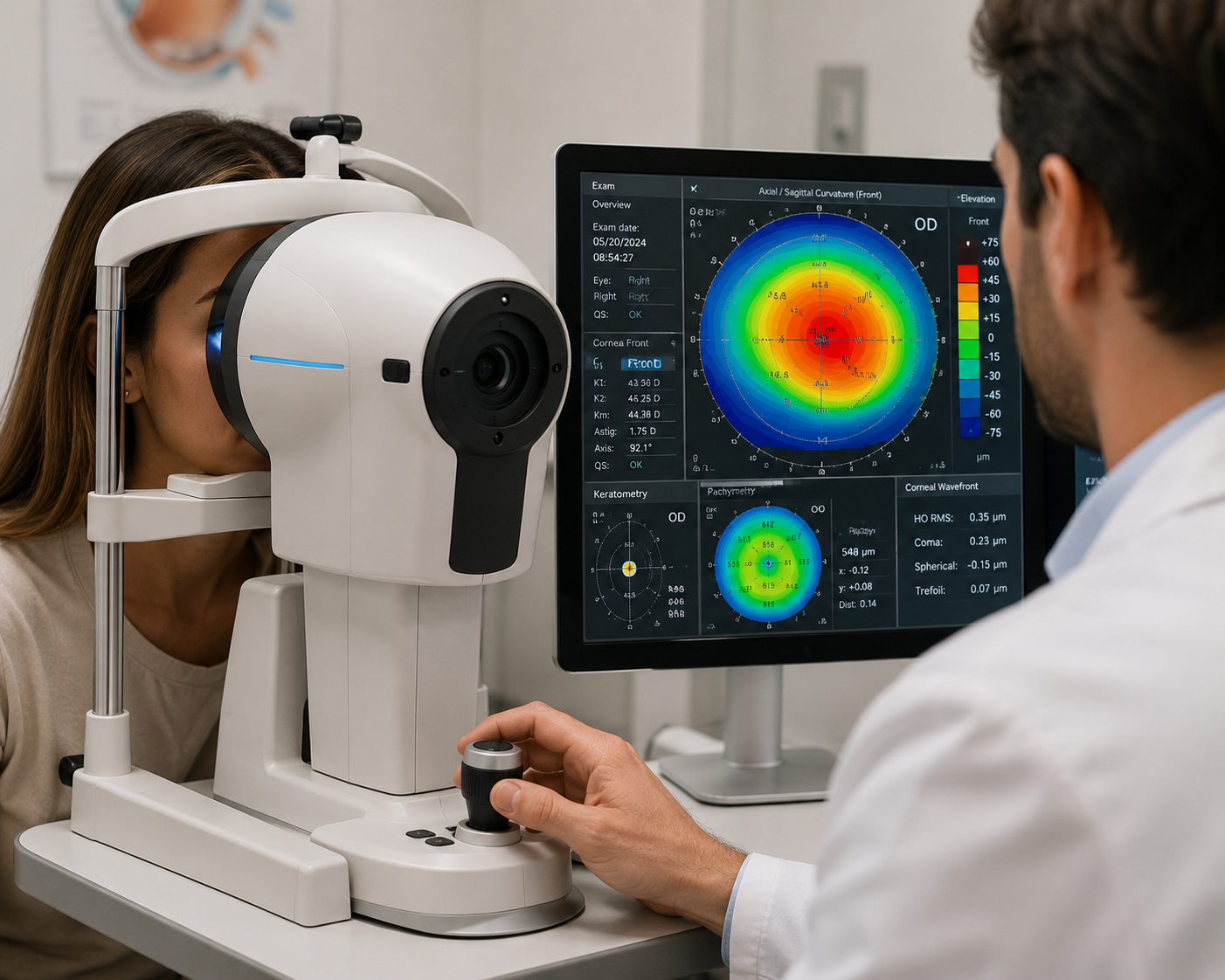
The base curve of the lens back surface is fitted to create the correct vault clearance, usually between 150 and 350 microns at the corneal apex, depending on the degree of irregularity. Get it too shallow and the lens will touch the cornea, causing abrasion. Get it too steep and the reservoir becomes so large that visual distortion and fogging become problems. This precision fitting is why providers at the home page of Texas Contact Lens Institute use corneal topography and optical coherence tomography (OCT) to map the eye before even selecting a trial lens.
RGP Material, Oxygen Transmission, and Why Lens Material Matters for Vision
The rigid material is not just about optics. It also determines how safely you can wear these lenses for the hours required. Gas-permeable lenses are made from silicone-acrylate or fluorosilicone-acrylate polymers with Dk values (oxygen permeability ratings) ranging from 100 to over 180 Dk/t in modern designs. The FDA classifies RGP lenses separately from soft lenses due to their distinct material properties and fitting requirements.
A higher Dk value means more oxygen reaches the corneal epithelium even though the lens does not directly touch the cornea. This matters because a hypoxic cornea becomes edematous and cloudy, directly degrading visual acuity. The combination of high-Dk material and the thin saline layer keeps oxygen flux sufficient for daily wear of 8 to 12 hours for most patients.

Advanced Optical Technologies: Wavefront, Toric, and Multifocal Designs
Modern scleral lens engineering goes well beyond basic spherical correction. If you have significant astigmatism layered on top of corneal irregularity, a toric scleral lens incorporates a cylindrical correction zone into the front optical surface. Because the lens is stable on the sclera and does not rotate with blinking the way a soft toric lens does, the cylindrical axis stays precisely positioned. That stability is one of the key advantages scleral lenses hold over soft lenses for complex prescriptions.
Wavefront-guided scleral lenses take this further. Aberrometry instruments measure not just sphere and cylinder but higher-order aberrations including coma, trefoil, and spherical aberration. These aberrations are the reason some patients with keratoconus describe vision as a smear of light even when corrected with standard lenses. By mapping and incorporating corrections for these higher-order errors directly into the lens optics, wavefront-guided designs can deliver significantly sharper acuity. Research published through the National Institutes of Health has demonstrated measurable improvements in high-contrast and low-contrast visual acuity with aberration-correcting scleral designs compared to standard spherocylindrical correction.
For patients over 40 managing presbyopia alongside corneal disease, multifocal scleral lenses incorporate alternating distance and near zones. The fitting complexity increases substantially with these designs, which is why working with a specialist, as noted on the meet the doctor page at Texas Contact Lens Institute, makes a measurable difference in outcomes.
Advantages, Drawbacks, and Who Benefits Most
Advantages:
Superior visual acuity for irregular corneas compared to glasses or soft lenses
Stable lens position due to scleral landing, reducing blur from lens movement
Continuous hydration from the fluid reservoir benefits patients with dry eye
Non-surgical alternative to corneal transplant in many keratoconus cases
Compatible with complex prescriptions including toric and multifocal corrections
Drawbacks:
Higher cost, often ranging from $1,500 to $4,000 per pair including fitting fees
Multiple fitting visits required before the final lens is finalized
Insertion and removal technique takes practice and patience
Not covered by all insurance plans; check your benefits carefully
For patients being referred by another provider, the referrals process at Texas Contact Lens Institute is straightforward and designed to keep your existing care team in the loop. The Mayo Clinic also notes that scleral lenses are frequently the most effective non-surgical management option for keratoconus before a transplant is considered.
Things to Know
The saline solution used in the reservoir must be preservative-free; standard saline can cause corneal inflammation over time.
Most scleral lenses last 1 to 3 years, though surface deposits and prescription changes are the most common reasons for earlier replacement.
You should never let the lens run dry during insertion; air bubbles under the lens cause significant visual disruption.
Wearing time beyond 12 to 14 hours daily increases hypoxia risk even with high-Dk materials.
Fitting scleral lenses for both eyes typically requires separate evaluations since each eye's topography differs.
Frequently Asked Questions
Q: How is a scleral lens different from a regular gas-permeable lens optically?
A standard RGP lens sits on the cornea and relies on the tear film to fill minor gaps, while a scleral lens creates a controlled fluid vault that fully replaces the corneal optical surface.
This distinction makes scleral lenses far more effective for irregular corneas where the tear film alone cannot compensate for the distortion. The engineered saline layer provides a consistent, optically smooth interface regardless of the corneal shape underneath.
Q: Can scleral lenses correct astigmatism caused by corneal irregularity?
Yes, the saline reservoir neutralizes corneal astigmatism, and toric front surface designs can address residual refractive astigmatism on top of that.
This dual-layer correction is one reason scleral lenses outperform soft torics in patients with both irregular and regular astigmatism. The stable fit ensures the cylinder axis does not shift with eye movement.
Q: How long can you wear scleral lenses each day?
Most patients wear scleral lenses comfortably for 8 to 14 hours per day, though individual oxygen tolerance and lens Dk values influence the upper limit.
Your fitter will typically start you at 6 to 8 hours and assess corneal health before extending wear time. Redness, blurred vision, or discomfort after lens removal are signs you may be exceeding your safe daily duration.
Q: Do scleral lenses work for presbyopia?
Yes, multifocal scleral lens designs are available and can correct both near and distance vision simultaneously.
These are more complex to fit than standard scleral lenses and require additional trial lens evaluations. They are particularly useful for patients over 40 who also have keratoconus or other corneal irregularities.
Q: What happens if the saline reservoir has an air bubble?
An air bubble under the lens creates a localized gap where the optical smoothing effect is lost, causing a dark or blurry spot in your visual field.
The fix is to remove the lens, refill it completely with fresh saline ensuring no air is trapped, and reinsert carefully. Proper technique during insertion eliminates this issue in most cases.
The Bottom Line on How Scleral Lens Optics Work
The optical engineering behind scleral lenses is precise and clinically powerful. The combination of a rigid refracting surface, a saline tear lens that neutralizes corneal irregularity, and stable scleral landing creates a visual correction system that glasses and soft lenses simply cannot replicate for complex corneal conditions. Understanding how does optics of a scleral lens work positions you to have a much more productive conversation with your fitter about vault depth, material selection, and design options.
If you are ready to explore whether scleral lenses are right for your eyes, the team at Texas Contact Lens Institute, located at 6020 W Parker Rd Ste 260, Plano, TX 75093, specializes in specialty contact lens fitting with a 5.0 rating across 12 reviews. You can visit the blog for more clinical insights or use the contact page to schedule your evaluation at (972) 299-8675.
