
Best Hygiene Practices with Scleral Lens Wear: Step-by-Step Protocols That Actually Work
Proper hygiene is the single most important factor in safe scleral lens wear. Following a consistent, evidence-based routine dramatically reduces your risk of serious eye infections, including corneal ulcers and sight-threatening conditions.
Key Takeaways
Always wash hands with mild, fragrance-free soap before handling scleral lenses.
Use a hydrogen peroxide-based solution for at least 6 hours to fully disinfect lenses.
Never rinse lenses or fill the bowl with tap water, as it can harbor Acanthamoeba.
Replace your lens case every 1-3 months and clean it daily to reduce contamination risk.
Protein and lipid deposits build up faster on scleral lenses due to the moisture chamber design — a daily surfactant cleaner is essential.
Schedule regular professional check-ins to catch fit or hygiene issues early.
Why Hand Hygiene Is the Starting Point for Every Scleral Lens Session
Before you touch your lenses, your hands must be completely clean. Wash with a mild, fragrance-free, lotion-free soap for at least 20 seconds, then rinse thoroughly and dry with a lint-free towel. Residue from moisturizers, hairspray, sunscreen, or scented soaps can transfer onto the lens surface and directly into the fluid reservoir that sits against your cornea.
This matters more with scleral lenses than with soft lenses because the bowl of saline solution that fills the lens creates direct, prolonged contact with the eye's surface. Any contamination you introduce during handling does not simply evaporate; it sits on your cornea for hours. According to research reviewed by the American Academy of Ophthalmology, approximately 99% of contact lens wearers report at least one hygiene risk behavior, making this the most common entry point for infection that you can learn more about when you visit the home page.

Daily Cleaning and Disinfection: The Hydrogen Peroxide Method
The most clinically supported approach to scleral lens disinfection uses a hydrogen peroxide-based solution, and it requires a minimum soak time of 6 hours. This is not a shortcut-friendly process. The peroxide must fully neutralize before the lens touches your eye. Skipping or shortening the neutralization cycle causes chemical burns to the cornea.
A complete daily care regimen for scleral lens wearers typically includes three steps:
Daily surfactant cleaner: Rub each lens for 1-2 minutes with a dedicated cleaning solution to physically remove protein, lipid, and mucin deposits. Clinical data shows that 62% of scleral lens patients use a separate daily surfactant cleaner, and those who skip it tend to accumulate more deposits.
Hydrogen peroxide disinfection: Submerge the lens in a peroxide system (such as Clear Care) for at least 6 hours, allowing full antimicrobial activity against bacteria, fungi, and protozoa.
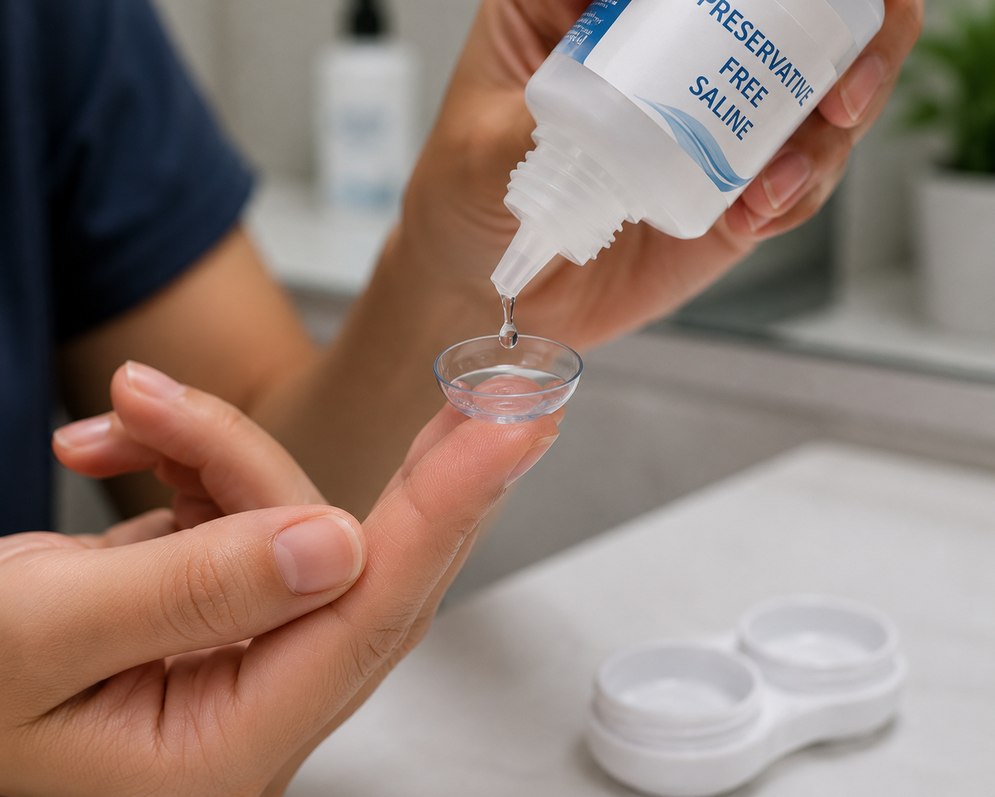
Preservative-free saline rinse: Before insertion, rinse lenses thoroughly with a sterile, preservative-free saline or dedicated filling solution. Never use multipurpose soft lens solution, tap water, or distilled water as a substitute.
For patients managing conditions like keratoconus or severe dry eye, theNIH National Eye Institute provides helpful background on why specialty lens hygiene carries elevated importance when discussed with specialists like those featured on themeet the doctor page.
Solution Management: What You Use and When You Use It
Every day means fresh solution, no exceptions. Topping off existing solution in your case dilutes the disinfectant concentration and significantly reduces its effectiveness. Empty your case completely, rinse it with fresh solution (not water), and let it air dry face-down on a clean tissue.
The danger of tap water cannot be overstated. Acanthamoeba, a microscopic organism found in municipal water supplies, swimming pools, and even well water, can adhere to contact lenses and cause Acanthamoeba keratitis, a painful and difficult-to-treat infection. The CDC's contact lens health resources outline this risk in detail and confirm that tap water exposure is one of the leading preventable causes of lens-related eye infections that you can discuss further via the contact page.
Single-use vials of non preserved saline are the gold standard for filling the scleral lens bowl before insertion. Preserved saline can cause ocular surface irritation with repeated daily exposure.
Lens Case Care and Replacement Timeline
Most lens wearers overlook the case itself. Research indicates that up to 81% of lens cases show contamination, with cases older than 9 months carrying the highest contamination rates. Your recommended routine:
Rinse the case with fresh disinfecting solution after each use (never tap water)
Rub the inside of the case with a clean finger to remove biofilm
Air dry face-down on a lint-free surface with the caps off
Replace the case every 1-3 months, or immediately if you notice discoloration, deposits, or visible damage

Protein and Deposit Management for Scleral Lens Wearers
This is where scleral lenses differ from standard soft or rigid gas permeable (RGP) lenses in an important way. The moisture chamber design means that tears, mucin, and proteins are constantly in contact with the inner bowl of the lens. Lipid and protein deposits accumulate faster as a result, affecting optical clarity and comfort.
Weekly enzyme treatments or more frequent surfactant cleaning cycles are often recommended for heavy depositors. If you notice fogging or reduced vision quality midday, deposits may be the cause. This is a conversation worth having with your specialist, and you can explore theservices page at Texas Contact Lens Institute to review the specialty fitting options and lens care guidance available.
Insertion, Removal, and the Tools That Help
Scleral lenses require a plunger-style insertion tool and a DMV scleral vial or similar filling device. Keeping these tools clean is part of your hygiene protocol. Rinse your plunger with fresh saline before and after each use, and replace it every 3-6 months.
Avoid inserting or removing lenses over a hard surface. A folded clean towel placed in your sink creates a safer landing zone if the lens is accidentally dropped. Do not insert lenses in the shower or near running water.
You should also remove lenses before swimming in any body of water, including pools, hot tubs, lakes, and oceans. Waterborne pathogens and chlorine residue both pose risks to lens integrity and eye safety.
Recognizing Contamination and Knowing When to Seek Help
Infection warning signs include increased redness, light sensitivity, unusual discharge, blurred vision, or significant pain. If any of these appear, remove your lenses immediately and contact your eye care provider without delay. Do not attempt to self-diagnose.
The Mayo Clinic's guide on contact lens-related eye infections outlines the full symptom picture and reinforces when professional evaluation is urgent and should be discussed in detail through the blog.
Texas Contact Lens Institute, located at 6020 W Parker Rd Ste 260, Plano, TX 75093, specializes in scleral lens fitting, keratoconus management, orthokeratology, myopia management, dry eye treatment, and specialty contact lens fitting. Their team maintains a 5.0 rating across patient reviews, and you can learn more by visiting the meet the doctor page or reaching out through the contact page directly.

Things to Know
Smoking is a documented risk factor for contact lens-related infections — quitting reduces this risk meaningfully.
Travel hygiene requires extra planning: bring single-use saline vials, a clean spare case, and avoid filling lenses with hotel tap water.
Preservative-free drops, not regular artificial tears, should be used while wearing scleral lenses to avoid compatibility issues.
If you wear scleral lenses due to a referral from another provider, Texas Contact Lens Institute accepts referrals and has a streamlined intake process.
Lens cases 9 months or older carry the highest contamination rates, making timely replacement non-negotiable.
Frequently Asked Questions
Q: Can I rinse my scleral lenses with tap water in an emergency?
No — tap water should never contact scleral lenses under any circumstances.
Tap water contains Acanthamoeba and other pathogens that can adhere to the lens and cause severe, vision-threatening infections. Carry a spare vial of preservative-free saline when traveling as your backup option.
Q: How often should I replace my scleral lens case?
Replace your lens case every 1 to 3 months, or sooner if it shows any visible contamination.
Lens cases are among the most overlooked hygiene factors. Biofilm builds up inside the case walls even with daily rinsing, and older cases have measurably higher contamination rates.
Q: Why do my scleral lenses fog up during the day?
Midday fogging is most commonly caused by lipid or mucin deposits entering the moisture chamber.
This can result from incomplete daily cleaning, a poorly fitting lens, or underlying dry eye. Discuss the issue with your specialist, as a fitting adjustment or cleaning protocol change often resolves it.
Q: Is hydrogen peroxide disinfection really necessary, or can I use multipurpose solution?
Hydrogen peroxide systems provide deeper disinfection and are strongly preferred for scleral lenses.
Multipurpose solutions are formulated for soft lenses and may not offer adequate antimicrobial activity for rigid scleral materials. They can also leave preservative residue that irritates the eye through the prolonged scleral lens moisture chamber.
Q: How do I know if my scleral lens hygiene routine is working?
Comfortable, clear vision with no redness, discharge, or sensitivity suggests your routine is effective.
Regular check-in appointments are the most reliable confirmation. Your provider can inspect lens surfaces for deposit buildup and assess the ocular surface for early signs of irritation before symptoms escalate.
The Bottom Line on Best Hygiene Practices with Scleral Lens Wear
Consistency is what separates safe scleral lens wear from risky wear. Every step in your routine, from washing your hands to replacing your case on schedule, adds a layer of protection that keeps your cornea healthy and your vision clear. Shortcuts in any part of this chain create real, preventable risk.
Your next step is to confirm your current routine with a qualified scleral lens specialist. Visit the home page or browse the blog at Texas Contact Lens Institute for additional patient resources. You can reach their Plano, TX office at +1 972-299-8675 to schedule a hygiene review or fitting consultation today.
Inserting and Removing Scleral Lenses Without Plungers: Finger Techniques That Actually Work
Knowing how to insert and remove scleral lenses without plungers is a skill every scleral lens wearer should have in their back pocket. These finger-based techniques work reliably in a pinch, whether you left your plunger at home, it cracked during travel, or you simply want a more tactile method of handling your lenses.
Key Takeaways
You can safely insert and remove scleral lenses without a plunger using proper finger positioning and lubrication.
Lubricate your eye with preservative-free saline or rewetting drops before any removal attempt to reduce friction and protect your cornea.
Keep your fingernails trimmed short and smooth before attempting any manual technique.
The lid margin technique is the most effective plunger-free removal method for most scleral lens wearers.
If your eye becomes red, painful, or increasingly irritated, stop immediately and contact your eye care provider.
Professional fitting and training from a qualified optometrist significantly improves your success rate with these techniques.
What Scleral Lenses Are and Why Handling Them Differs From Standard Contacts
Scleral lenses are large-diameter rigid gas-permeable lenses, typically ranging from 14.5 mm to over 22 mm in diameter, that vault entirely over the cornea and rest on the white part of the eye called the sclera. Unlike soft contacts, they create a fluid-filled reservoir between the lens surface and the corneal tissue. This reservoir provides consistent hydration and a smooth optical surface, making sclerals the preferred option for people with keratoconus, severe dry eye, corneal scarring, and other irregular cornea conditions.
Because of their size and the vacuum seal they form on the eye, scleral lenses require more deliberate handling than standard soft lenses. Plungers (also called DMV tools) are the conventional removal aid, but they are not always available, which is why understanding how to handle these lenses manually on the home page of Texas Contact Lens Institute protects your eye health during travel, emergencies, or situations where your equipment is unavailable.

Things to Know
Scleral lenses form a mild vacuum seal against the sclera, which is what makes removal feel more resistant than removing a soft lens.
Preservative-free saline is the only fluid recommended to fill the lens bowl before insertion; tap water is never safe for contact lens use.
The CDC guidelines on contact lens hygiene apply to scleral lenses too, including thorough handwashing before any handling.
If your lens has a toric or prism ballast design for astigmatism correction, it must be rotated to the correct orientation before insertion.
Not all optometrists fit scleral lenses, and you should seek a provider with demonstrated specialty contact lens experience, such as the team you can meet the doctor with at Texas Contact Lens Institute in Plano, TX.
Scleral lens handling techniques can vary slightly depending on lens diameter and your personal eye anatomy, so individualized training from your fitter matters.
Preparation Steps Before Inserting or Removing Scleral Lenses
Preparation is not optional. Rushing into insertion or removal without the right setup increases your risk of dropping the lens, scratching your cornea, or contaminating the fluid reservoir.
Handwashing and Nail Care
Wash your hands thoroughly with soap and water for at least 20 seconds, then dry them with a lint-free towel. According to the American Optometric Association, improper hand hygiene is one of the leading contributors to contact lens-related eye infections. Check your fingernails before every session. They should be trimmed short and filed smooth. Even a slight edge can scratch the cornea or tear the lens during manual handling.
Setting Up Your Workspace
Work over a clean, flat surface or a folded towel to cushion the lens if it drops. Use a well-lit space or a magnifying mirror at counter height to avoid leaning over a sink. Have your preservative-free saline solution, a clean lens case, and your lens cleaning solution nearby before you begin.
Lubricating the Eye First
Before any removal attempt, instill two to three drops of preservative-free saline or rewetting drops into the eye. This step reduces friction between the lens edge and the conjunctival tissue, making both the lid margin technique and the pinch method more comfortable and less likely to cause irritation. You can find guidance on recommended eye drops and compatible solutions through the services offered at Texas Contact Lens Institute.
How to Insert Scleral Lenses Without a Plunger
The standard insertion method already relies primarily on the fingers rather than a plunger, so this is one area where most wearers are already comfortable. That said, there are specific details that make finger insertion more reliable.
The Three-Finger Hold
Place the lens on the tips of your thumb, index finger, and middle finger, forming a tripod support. This distributes the weight of the lens evenly and reduces the chance of tipping or spilling the saline during the process. Fill the bowl completely with preservative-free saline. The lens should be full to the brim with no air bubbles visible, which can cause blurry vision or discomfort after insertion.
Tilting and Opening the Lids
Tilt your head downward so you are looking at the floor, not straight ahead. Using your non-dominant hand, use one finger to hold the upper eyelid open against the brow bone and another finger or your thumb to hold the lower lid down. Bring the filled lens up directly under your eye, keeping it level to avoid spilling. When the lens contacts the eye, hold it in place for two to three seconds before slowly releasing your eyelids to capture it in position.
Centering the Lens
After releasing your lids, keep your head tilted down for a moment, then slowly raise it to look straight ahead. Gently blink. If vision is clear and comfortable, the lens is centered correctly. If you notice haziness, there may be an air bubble trapped beneath the lens, and you will need to remove and reinsert it.
How to Remove Scleral Lenses Without a Plunger
This is where most wearers feel uncertain without a DMV tool. Two primary finger techniques work well, and both are safe when performed correctly, as detailed in the blog at Texas Contact Lens Institute.
The Lid Margin Technique
This is widely considered the most effective method for removing scleral lenses without a plunger. Here is how to do it step by step:
Wash and dry your hands thoroughly.
Instill preservative-free drops to lubricate the eye.
Look straight ahead into a mirror.
Place the index finger of your dominant hand at the outer corner of your eye, at the junction where the upper and lower lids meet.
Gently pull the outer lid corner slightly toward your ear to create tension.
Blink deliberately and firmly. The tension at the lid margin creates a break in the vacuum seal, and the lens will pop forward onto your lower lid or into your waiting cupped hand.
Practice this over a folded towel or a flat surface until you develop confidence in the motion.
The Pinch and Slide Technique
The pinch method uses the index finger and thumb of your dominant hand. Look upward. Place the pad of your index finger on the lower edge of the lens, which will be exposed below your lower lid when you look up. Very slowly slide the lens downward, off the cornea and onto the sclera. Once it has moved down, gently pinch the lens edge between your thumb and index finger to lift it away from the eye. This technique requires patience. Do not pull quickly. A slow, deliberate slide breaks the vacuum more safely than a sharp tug.
Using a Sterile Q-Tip or Gauze as Assistance
If your fingertip alone does not provide enough leverage at the lens edge, a sterile cotton swab or small piece of clean gauze can help. Apply gentle pressure at the lower margin of the lens to help tilt it forward and break the seal. The soft material provides better grip on the lens edge than a bare fingertip, especially if you have shorter fingers or difficulty reaching the lens margin. This technique is a legitimate backup, though it should never replace proper training from a qualified specialist.
Safety Guidelines and When to Stop
Even gentle manual techniques carry some risk if repeated without success. The American Academy of Ophthalmology recommends stopping any contact lens removal attempt that causes escalating pain, redness, or tearing, and seeking professional help instead.
If the lens has not come out after three or four careful attempts, do not continue forcing it. Excessive pressure or repeated failed attempts raise the risk of corneal abrasion or conjunctival damage. Instead, instill more rewetting drops to soften the seal further, rest your eye for a few minutes, and try once more calmly. If your eye becomes noticeably red, painful, or significantly irritated, stop all attempts and call your eye care provider.
Texas Contact Lens Institute, located at 6020 W Parker Rd Ste 260, Plano, TX 75093 (phone: 972-299-8075), specializes in scleral lens fitting and patient education. Their referrals page can direct new patients or transfer cases to the right specialist. You can also learn more about their approach to specialty lens care through the contact page to schedule a consultation.
Building Long-Term Confidence With Scleral Lens Handling
Learning how to insert and remove scleral lens without plungers is a skill that improves with deliberate practice. Most new wearers feel awkward during the first few weeks, and that is completely normal. The Mayo Clinic's overview of contact lens safety highlights that most lens-related complications stem from improper handling rather than the lenses themselves.
Reading about techniques is a starting point, but in-office training is where lasting competence comes from. The meet the doctor page at Texas Contact Lens Institute gives you a clear picture of who will be guiding your fitting and training process. Their blog also covers ongoing tips and updates relevant to specialty lens wearers.
If you have questions about your specific lens design, handling challenges, or are experiencing persistent difficulty, reach out directly through the contact page to schedule an appointment or ask about personalized coaching for lens insertion and removal.
Frequently Asked Questions
Q: Is it safe to remove a scleral lens without any tools at all?
Yes, finger-based techniques are safe when performed correctly with clean, trimmed hands and lubricated eyes.
Both the lid margin and pinch techniques are accepted methods used by scleral lens wearers worldwide. The key is to apply gentle, controlled pressure and stop if the eye becomes painful or red.
Q: What should I fill a scleral lens with if I run out of saline?
You should only use preservative-free saline solution to fill the lens bowl before insertion.
Tap water, distilled water, and standard saline with preservatives are not safe substitutes. CDC contact lens safety resources clearly state that tap water should never contact lenses or lens cases due to the risk of Acanthamoeba keratitis, a serious eye infection.
Q: Why does my scleral lens feel stuck and hard to remove?
The lens creates a mild vacuum seal against the sclera, which can feel stronger if the eye is dry or the lens has been worn for many hours.
Instilling rewetting drops before removal usually resolves this. If the lens consistently feels stuck, discuss your wearing schedule and fit with your eye care provider.
Q: How long does it take to get comfortable inserting and removing scleral lenses without a plunger?
Most wearers develop a reliable routine within two to four weeks of consistent daily practice.
Early sessions may take 15 to 20 minutes. With repetition and any adjustments suggested by your fitter, the process typically shortens to five minutes or less.
Q: Can I use any type of cotton swab to help remove my scleral lens?
Only sterile, individually wrapped cotton swabs should be used near the eye.
Avoid swabs that have been sitting open, as they can carry dust or bacteria. The cotton swab method is a backup technique and should not become a daily habit without guidance from your eye care provider.
The Bottom Line on How to Insert and Remove Scleral Lens Without Plungers
Finger-based insertion and removal techniques are reliable, safe, and worth mastering regardless of whether you always carry a plunger. The lid margin and pinch techniques give you real independence in managing your lenses, and proper preparation including handwashing, nail care, and lubrication dramatically improves your success rate.
If you are based in the Dallas-Fort Worth area and want personalized instruction from a rated specialist, Texas Contact Lens Institute in Plano, TX offers scleral lens fitting, training, and ongoing support. Reach out today to schedule your consultation and get the hands-on guidance that makes all the difference.
Natural Relief for Dry Eyes: Simple Lifestyle Hacks and the Truth About Homeopathic Drops
Dry eyes can be managed at home through targeted lifestyle changes like adjusting screen habits, improving hydration, and modifying your environment. At the same time, the truth about homeopathic drops is more complicated than most product labels suggest, and knowing the difference between helpful and harmful options can protect your vision.
Key Takeaways
Approximately 16 million Americans have diagnosed dry eye disease, but prevalence estimates suggest up to 50% of all adults experience symptoms at some point.
The 20-20-20 rule, increased water intake, and omega-3-rich foods are among the most evidence-backed lifestyle strategies.
Homeopathic eye drops are not proven effective by rigorous scientific standards, and several products have been recalled due to bacterial contamination.
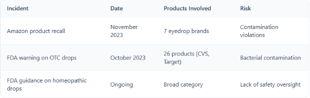
The FDA has issued warnings on 26 over-the-counter drops sold at major US retailers including CVS and Target due to contamination risks.
Artificial tears backed by clinical evidence remain the first-line OTC recommendation for most dry eye sufferers.
Persistent or worsening symptoms require a professional evaluation, not a trip to the supplement aisle.
Why So Many Americans Are Dealing with Dry Eyes Right Now
Dry eye disease is no longer a niche complaint. According to available prevalence data, approximately 16 million Americans carry a formal diagnosis, and broader estimates suggest up to 50% of adults experience symptoms at some point. Among adults over 40, between 10% and 20% report moderate to severe issues.
Several factors are driving this trend upward. Digital device usage sits at the top of the list. When you stare at a screen, your blink rate drops significantly, which means the tear film over your eye dries out faster than it can be replenished. Researchers are increasingly concerned about rising rates of dry eye symptoms even in children due to early and heavy screen exposure.
Other contributing factors include:
Contact lens wear, particularly older lens types not optimized for oxygen permeability
Living or working in air-conditioned or heated environments with low humidity
Aging, which reduces tear production naturally
Certain medications, including antihistamines, antidepressants, and blood pressure drugs
Autoimmune conditions like Sjögren's syndrome and rheumatoid arthritis
Understanding what's behind your symptoms is the first step before reaching for any remedy, whether it's a lifestyle change or a product off a store shelf.

Proven Lifestyle Hacks That Actually Reduce Dry Eye Symptoms
Before you buy anything, several free and low-cost adjustments can make a measurable difference in how your eyes feel throughout the day.
Hydration and Diet Changes Worth Making
Your tear film is roughly 98% water, so chronic mild dehydration directly affects tear volume and stability. Drinking adequate water throughout the day, typically aiming for around 8 glasses (64 oz), is a baseline that many dry eye sufferers overlook.
Diet plays a supporting role as well. Fatty fish like salmon, mackerel, and tuna are rich in omega-3 fatty acids, which improve the oily (lipid) layer of the tear film. That lipid layer is what slows evaporation. Without it, even a normal volume of tears evaporates too quickly, leaving the eye surface exposed. TheAmerican Academy of Ophthalmology recommends omega-3 supplementation as a reasonable supportive measure for dry eye management.
Foods to add to your routine:
Salmon, sardines, and mackerel (2-3 servings per week)
Flaxseed and chia seeds for plant-based omega-3s
Leafy greens rich in vitamin A, which supports healthy corneal surface cells
Walnuts as a convenient omega-3 snack option
Screen Time Habits and the 20-20-20 Rule
One of the simplest, most consistently recommended strategies is the 20-20-20 rule. Every 20 minutes, look at something at least 20 feet away for at least 20 seconds. This forces a natural blink reset and gives your eyes a brief recovery period.
Additional screen hygiene practices worth adopting:
Position your monitor slightly below eye level, which reduces the exposed eye surface area
Reduce screen brightness to match ambient room lighting
Use a matte screen filter to cut glare
Set a reminder app or use built-in screen time tools on your phone or computer
Environmental Controls at Home and at Work
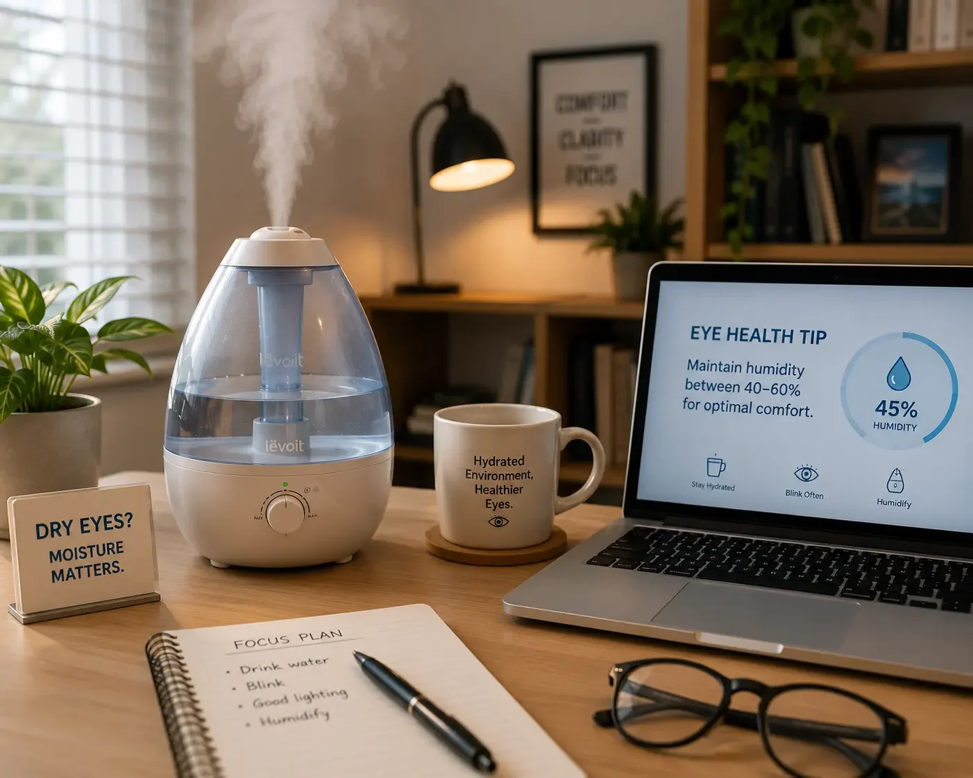
Low indoor humidity is a major but underappreciated trigger. Central heating in winter and air conditioning in summer can drop indoor humidity well below 40%, which accelerates tear evaporation. Investing in a quality humidifier for your bedroom or home office is one of the most cost-effective changes you can make. Units designed for room coverage typically run between $30 and $80.
If you wear contact lenses, switching to daily disposable lenses or specialty designs may also reduce surface irritation. Theservices offered at specialty clinics like Texas Contact Lens Institute include dry eye treatment and specialty contact lens fitting for patients who can't tolerate standard lenses.
The Real Truth About Homeopathic Eye Drops
Here's where it gets important. The term "natural" on an eye drop label does not mean safe, and "homeopathic" does not mean effective.
What Homeopathy Actually Claims and Why Scientists Reject It
Homeopathy is based on two core principles: the "law of similars" (substances that cause symptoms in healthy people can treat those same symptoms in sick people) and the "law of infinitesimals" (diluting a substance makes it more potent). Neither principle has been validated by modern science. TheNational Institutes of Health's National Center for Complementary and Integrative Health states clearly that the effectiveness of homeopathic substances has not been demonstrated by scientific research.
Some clinical trials have shown modest symptom relief from specific homeopathic formulas. A study comparing Homeoptic® to Larmabak® found that Homeoptic® provided greater relief for mild-to-moderate dry eye symptoms. However, this is a narrow finding on a single product, not a validation of homeopathy as a category.
FDA Warnings, Recalls, and the Contamination Risk You Need to Know
Eyes are what scientists call "immune-privileged" sites. The blood-eye barrier limits the immune system's access to ocular tissue, which means bacteria introduced through contaminated drops can cause serious, rapid infections including bacterial keratitis, a condition that can result in permanent vision loss if untreated.
Recent regulatory actions underscore how serious this risk is:
TheFDA's current policy on homeopathic drug products emphasizes that many homeopathic products have not been reviewed for safety or efficacy. The agency has been pushing for expanded oversight, including mandatory recall powers and manufacturer inspections.
If you're currently using a homeopathic eye drop, check the brand against available recall databases and verify the manufacturing location. Products made outside the US face fewer regulatory controls during production.
What to Use Instead
For most people with mild to moderate dry eye, preservative-free artificial tears are the evidence-backed OTC starting point. Look for products containing ingredients like carboxymethylcellulose or sodium hyaluronate. Gel drops work better for nighttime use when thicker lubrication is acceptable.
If you're unsure whether your symptoms are caused by meibomian gland dysfunction (evaporative dry eye) versus aqueous deficiency (low tear production), a professional evaluation will point you to the right approach.

When Lifestyle Hacks Aren't Enough: Recognizing the Signs You Need Professional Help
Lifestyle changes and over-the-counter drops are appropriate for mild, situational dry eye. But if your symptoms persist beyond two to three weeks of consistent self-care, or if you're experiencing any of the following, a professional evaluation is warranted:
Blurred vision that doesn't clear with blinking
Eye pain or significant burning not relieved by lubricating drops
Light sensitivity
Discharge or redness suggesting possible infection
Symptoms that worsen despite treatment
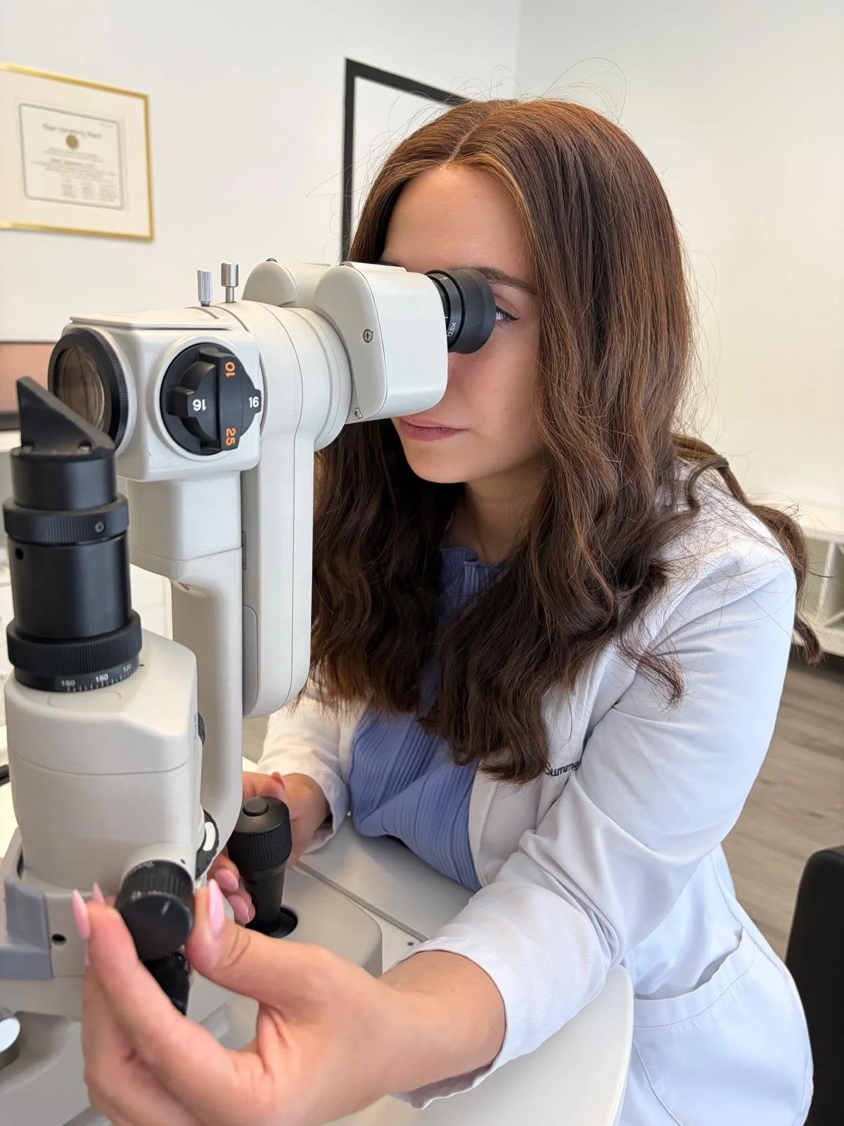
The team at Texas Contact Lens Institute, located at 6020 W Parker Rd Ste 260 in Plano, TX, specializes in dry eye treatment alongside scleral lens fitting for patients with more complex ocular surface conditions. You can reach them directly at +1 972-299-8675. Visiting themeet the doctor page gives you background on the clinical expertise available before your first appointment.
For patients whose dry eye symptoms are tied to keratoconus or corneal irregularities, scleral lenses can vault over the damaged surface entirely and provide both vision correction and a fluid reservoir that keeps the eye continuously lubricated. This is one area where a generalist eye care provider may not have the same depth of expertise as a specialty practice.
Thereferrals section of the Texas Contact Lens Institute website is designed specifically for cases where a referring physician wants to connect a patient with specialty dry eye care. If your optometrist or ophthalmologist has recommended a second opinion on your dry eye management, that pathway is available.
Things to Know
The 20-20-20 rule is widely cited but only effective if you actually follow it consistently. Setting a timer or using a dedicated app makes compliance much more realistic.
Omega-3 supplements show more consistent results in studies than dietary omega-3 alone because it's hard to eat enough fatty fish regularly to achieve therapeutic levels.
Not all artificial tears are equal. Preservative-containing drops are fine for occasional use but can worsen the eye surface if used more than four times daily.
Homeopathic products sold in the US before 1938 were grandfathered under the Food, Drug, and Cosmetic Act and have not undergone the same safety review as other OTC medications.
Wearing contact lenses for longer hours than prescribed is one of the most common and preventable causes of chronic dry eye symptoms.
TheMayo Clinic's dry eye overview and the American Academy of Ophthalmology are both reliable resources for verifying treatment claims you encounter online.
Frequently Asked Questions
Q: Can drinking more water actually improve dry eye symptoms?
Yes, staying well-hydrated supports tear film stability, though it works best as part of a broader approach.
Dehydration reduces the aqueous component of your tears, making them less effective at lubricating the eye surface. Aiming for 64 oz of water daily is a reasonable baseline, but pairing hydration with dietary omega-3s and environmental adjustments produces better results than water intake alone.
Q: Are there any homeopathic eye drops that are actually safe to use?
Some specific products have shown limited clinical benefit, but the category as a whole lacks rigorous safety oversight.
The FDA has issued repeated warnings about contamination risks in homeopathic drops, including bacterial contamination found in products at major US retailers. If you choose to use a homeopathic product, verify it hasn't been recalled and confirm it's manufactured in an FDA-inspected facility.
Q: How do I know if I have evaporative dry eye versus tear deficiency dry eye?
The two types require different treatments, and distinguishing them accurately typically requires a clinical evaluation with specific diagnostic tests.
Evaporative dry eye is usually caused by meibomian gland dysfunction, where the oily layer of the tear film breaks down. Aqueous deficiency dry eye involves reduced tear production. A specialist can run tests like meibography or tear osmolarity to identify which type you have.
Q: Is the 20-20-20 rule backed by clinical evidence?
Research supports it as a practical method for reducing digital eye strain and encouraging blink rate normalization.
While large-scale randomized controlled trials are limited, smaller studies and clinical recommendations from organizations like the American Academy of Ophthalmology support its use as a low-risk, cost-free habit for screen users.
Q: When should I stop trying home remedies and see a specialist?
If symptoms persist for more than two to three weeks despite consistent self-care, or if you experience pain, vision changes, or discharge, professional evaluation should not be delayed.
Delaying care for dry eye caused by an underlying condition can lead to corneal surface damage over time. Texas Contact Lens Institute offers dry eye treatment and specialty care; you can reach out through thecontact page or call +1 972-299-8675 to schedule an evaluation.
The Bottom Line on Natural Relief for Dry Eyes
Natural relief for dry eyes is genuinely achievable for many people through consistent lifestyle adjustments, including better hydration, omega-3 intake, screen habit changes, and environmental improvements. These strategies are low-risk, low-cost, and supported by clinical evidence. Homeopathic drops, however, occupy a different category. While isolated products have shown modest results, the category lacks the regulatory framework and scientific foundation to be recommended broadly, and contamination risks are real and documented.
If you've been managing symptoms on your own and aren't seeing improvement, the next step is a professional evaluation. You can start by browsing the blog at Texas Contact Lens Institute for clinically informed content on dry eye and specialty lens care, or visit the home page to learn more about their full range of services. For persistent or complex dry eye, specialty care isn't an overreaction. It's the most direct path to lasting relief.
Scleral Lenses: Who Actually Needs Them and What You Should Expect
Scleral lenses are large-diameter, rigid gas permeable contact lenses that vault over the cornea and rest on the whites of the eyes, making them the preferred option for people with irregular corneas or severe dry eye. Understanding why someone would need a scleral lens comes down to whether standard lenses or glasses fail to correct vision adequately or cause too much discomfort.
Key Takeaways
Scleral lenses are not standard contacts; they are a medical-grade solution for eyes that other correction methods can't address effectively.
Keratoconus, severe dry eye, post-surgical irregularities, and corneal scarring are the most common reasons people switch to sclerals.
The fluid reservoir between the lens and the cornea is a core feature, not a side benefit; it's what makes them work for both vision correction and surface healing.
Costs typically range from $1,500 to $4,000 per pair, and insurance coverage varies significantly by plan and diagnosis.
The fitting process requires a specialist and can take multiple appointments, but patient satisfaction rates are high once the right fit is achieved.
With proper care, scleral lenses last one to two years and most people adapt to insertion and removal within a few weeks.
What Scleral Lenses Actually Are (And How They Work)
A scleral lens looks nothing like the soft, thin discs most people picture when they think of contacts. These lenses measure between 14.5mm and 24mm in diameter, which is significantly larger than a standard soft lens (typically 14mm to 16mm). That extra size is the whole point.
Because of their diameter, scleral lenses completely vault over the cornea. They bridge across the entire corneal surface and land on the sclera, the firm white tissue surrounding the eye. The space created between the back of the lens and the front of the cornea fills with preservative-free saline solution, forming a liquid reservoir that keeps the corneal surface continuously hydrated throughout wear.
The lens itself is made from rigid gas permeable (RGP) material, which allows oxygen to pass through to the cornea. This was a historical limitation of earlier hard lenses, but modern RGP materials have largely eliminated that concern for most wearers.
The vaulting mechanism and fluid chamber are what separate sclerals from every other lens type. They don't touch the cornea at all during normal wear. That's the feature that makes them therapeutic for damaged or irregular corneal surfaces, not just a corrective tool.

Conditions That Make Scleral Lenses Medically Necessary
This is the core of why someone would need a scleral lens. The conditions below are the ones that most consistently push patients away from standard corrective options and toward sclerals.
Keratoconus and Other Corneal Ectasia Conditions
Keratoconus causes the cornea to progressively thin and bulge outward into a cone shape. That irregular surface means light doesn't refract consistently, and glasses or soft contact lenses can't neutralize the distortion. Scleral lenses vault over the cone entirely, using the fluid layer as an optical corrector.
Clinical data supports this strongly. Research has shown visual gains from a baseline logMAR of 0.50-0.53 down to post-treatment logMAR of 0.08-0.09 in keratoconus patients fitted with sclerals. That's a dramatic improvement in measurable visual acuity. Pellucid marginal degeneration and post-LASIK ectasia respond similarly.
Severe Dry Eye Disease
Severe dry eye is more than discomfort. For patients with aqueous-deficient or evaporative dry eye, wearing any contact lens can feel impossible. Scleral lenses change the equation because the saline reservoir sits between the lens and the eye constantly, acting as a synthetic tear film that doesn't evaporate.
In clinical studies of symptomatic soft lens wearers who switched to sclerals, 45% of participants reported wanting to continue scleral lens wear after just one month, citing significant symptom improvement.
Stevens-Johnson Syndrome and Graft vs. Host Disease
Both conditions can cause severe conjunctival scarring, lid abnormalities, and chronic ocular surface inflammation. The corneal surface in these patients is fragile, often producing almost no natural tears. The scleral lens protects the surface, delivers continuous hydration, and in some cases is the only way these patients can maintain functional vision without surgery.
Post-Surgical Complications
After LASIK, PRK, radial keratotomy, or corneal transplants, some patients develop irregular astigmatism that neither glasses nor soft lenses can correct. The corneal surface has been altered in ways that are no longer spherical or predictably curved. Scleral lenses neutralize that irregularity through the liquid-filled chamber.
Corneal Scarring and Trauma
Scarring from chemical burns, infections like acanthamoeba keratitis, or physical trauma to the eye creates irregular elevations and depressions in the corneal surface. A scleral lens effectively smooths over those irregularities optically while also protecting the damaged tissue.

Advantages That Set Scleral Lenses Apart From Other Options
The advantages are meaningful for the right patient. Vision correction through sclerals can address irregularities that are impossible to fix with any other non-surgical method. The lens doesn't shift on the eye because it rests on the stable sclera, not the curved cornea, so vision stays consistent throughout the day.
For patients with extreme sensitivity, the fact that the lens never contacts the cornea is not a minor perk. It's the difference between being able to function at work and not being able to open their eyes comfortably.
There is also a surgical avoidance factor. Many patients who would otherwise need a corneal transplant find that scleral lenses manage their condition well enough to delay or avoid surgery entirely. That's a significant quality-of-life and financial consideration.
Drawbacks and Practical Challenges to Anticipate
No lens type is without trade-offs, and sclerals have a genuine learning curve.
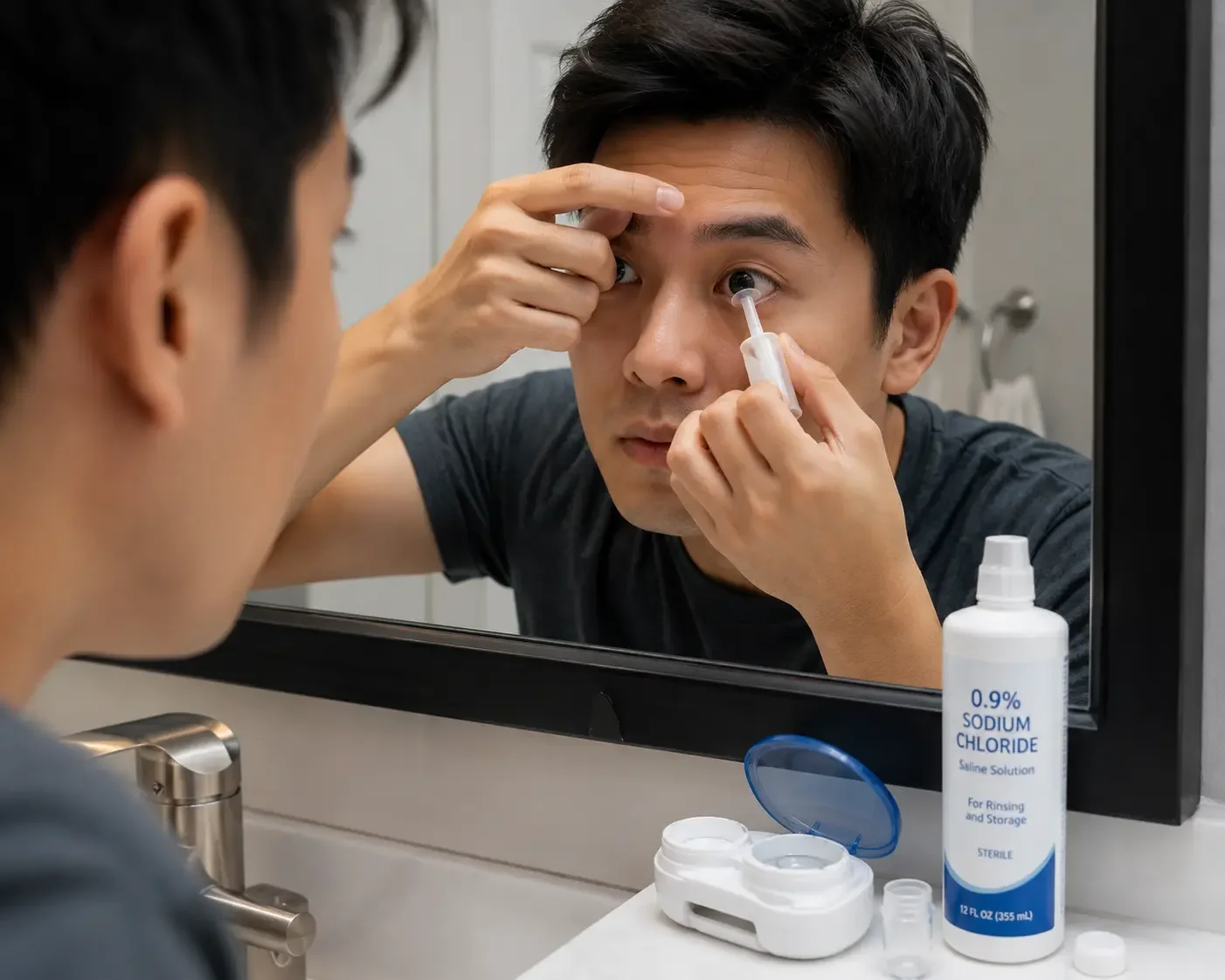
Insertion and removal require practice. You apply the lens while it's filled with saline, which means tilting the lens without spilling. Most people need two to three weeks before they feel comfortable.
The fitting process is lengthy. Getting the right fit requires multiple appointments with a specialist trained in complex contact lens fitting. Two to four visits before achieving an optimal fit is common.
Cost is a real barrier. At $1,500 to $4,000 per pair, scleral lenses are significantly more expensive than standard options. Insurance coverage through plans like vision plans often does not cover them. Medical insurance (for diagnoses like keratoconus) may contribute, but prior authorization is frequently required and not guaranteed.
Maintenance takes time. Lenses must be cleaned, stored in proper solution, and refilled with preservative-free saline every time you put them in. The routine is more involved than soft lens care.
Mid-day fogging can occur. Some patients experience clouding of the saline layer as the day progresses, requiring removal, cleaning, and reinsertion. Adjustments in fit or solution type can usually address this.
When You Should Contact Your Eye Doctor
Scleral lens wear is generally safe when managed properly, but certain symptoms require prompt attention.
Sudden blurry vision that doesn't clear after reinserting the lens
Eye redness that persists after lens removal
Pain, burning, or foreign body sensation during wear
Any visible change in the lens (chips, cracks, warping)
Signs of infection: discharge, excessive tearing, light sensitivity
I would not recommend trying to troubleshoot these symptoms on your own. Scleral lens-related complications can escalate quickly, especially for patients who already have compromised corneal tissue. Any of these signs warrant a call to your fitting specialist the same day.
How Long Scleral Lenses Last and What Daily Wear Looks Like
Scleral lenses typically last one to two years with proper care. The rigid material is more durable than soft lenses, but it scratches and deposits proteins over time, which can degrade optical clarity and increase the risk of eye irritation.
Daily wear duration varies by patient. Most practitioners recommend starting with four to six hours and gradually increasing wear time as the eye adapts. Many patients ultimately wear their lenses for eight to twelve hours a day. Some patients with severe dry eye or surface disease may have shorter tolerance windows, particularly early in wear.
The cleaning routine includes:
Rinsing the lens with multipurpose or hydrogen peroxide-based solution after removal
Cleaning with a daily cleaner or digital rubbing step to remove protein deposits
Storing overnight in appropriate solution
Rinsing with preservative-free saline before filling and reinserting each morning
Enzymes or protein-removing cleaners are sometimes added weekly for patients who deposit heavily. Your fitting specialist should outline a routine specific to your lens material and wear pattern.
Things to Know
Scleral lenses require fitting by a specialist with experience in complex lens fitting, not all optometrists or ophthalmologists have this training.
The saline used to fill the lens must be preservative-free; standard saline with preservatives can cause corneal toxicity with sclerals.
Medicare may cover scleral lens fittings under certain diagnoses, but coverage rules changed in recent years and vary by contractor.
Not every patient with keratoconus needs sclerals immediately; milder cases often do well with soft lenses or corneal RGP lenses first.
Children and teenagers with keratoconus are increasingly being fitted with sclerals, particularly when the condition progresses rapidly.
The fitting process involves corneal topography, optical coherence tomography (OCT), and often a trial set of diagnostic lenses before final ordering.

Make it stand out
Whatever it is, the way you tell your story online can make all the difference.
Frequently Asked Questions
Q: How much do scleral lenses cost in the United States?
Scleral lenses typically cost between $1,500 and $4,000 per pair, not including fitting fees, which can add $200 to $500 or more.
Vision insurance rarely covers the full cost. If you have a diagnosed condition like keratoconus, your medical insurance may cover a portion under durable medical equipment or specialty contact lens benefits, but you'll likely need documentation from your eye doctor and prior authorization.
Q: Are scleral lenses safe to sleep in?
No, scleral lenses are not approved for overnight wear and should be removed before sleeping.
Sleeping in any contact lens significantly increases infection risk by reducing corneal oxygenation. With sclerals, the risk is compounded by the fact that the fluid reservoir can become a breeding ground for bacteria if the lens stays in for extended periods. Remove, clean, and store them nightly.
Q: How do I know if I'm a good candidate for scleral lenses?
You're likely a good candidate if you have an irregular cornea, chronic dry eye, or a condition like keratoconus that standard lenses can't correct adequately.
The best way to find out is a consultation with a specialist who performs corneal topography and assesses your ocular surface. They'll evaluate your corneal shape, tear production, and current prescription to determine whether sclerals are the most appropriate option.
Q: How long does it take to get used to scleral lenses?
Most people adapt to the physical sensation within two to four weeks, though mastering insertion and removal can take longer for some patients.
The lens itself feels different from soft lenses because of its rigidity and size. Many patients report that after the initial adaptation period, sclerals feel more comfortable than soft lenses because they don't touch the sensitive corneal surface.
Q: Can children wear scleral lenses?
Yes, children, including teenagers with progressing keratoconus, are fitted with scleral lenses when their condition warrants it.
The key considerations for younger patients are their ability to handle insertion and removal independently, which most teenagers can learn with practice. Parental involvement during the fitting and learning process is strongly encouraged, and more frequent follow-up appointments are typically scheduled to monitor corneal changes.
The Bottom Line on Why Someone Would Need a Scleral Lens
Scleral lenses exist for one core reason: to give people with eyes that conventional correction methods can't adequately serve a real solution. Whether the issue is a progressive corneal disease, a damaged ocular surface, post-surgical irregularity, or severe dry eye, sclerals address the problem from the outside in, protecting the eye while simultaneously correcting vision to a level that most patients haven't experienced with any other option.
If you've been told your eyes are "too difficult to fit," or if glasses and soft lenses have never worked well for you, a consultation with a specialist trained in scleral lens fitting is a worthwhile next step. Ask your optometrist or ophthalmologist for a referral to a contact lens specialist, or use the American Academy of Optometry's directory at aaopt.org to find a credentialed fitter in your area.

Redefine Success
It All Begins Here
Confidence doesn’t always arrive with a bold entrance. Sometimes, it builds quietly, step by step, as we show up for ourselves day after day. It grows when we choose to try, even when we’re unsure of the outcome. Every time you take action despite self-doubt, you reinforce the belief that you’re capable. Confidence isn’t about having all the answers — it’s about trusting that you can figure it out along the way.
The key to making things happen isn’t waiting for the perfect moment; it’s starting with what you have, where you are. Big goals can feel overwhelming when viewed all at once, but momentum builds through small, consistent action. Whether you’re working toward a personal milestone or a professional dream, progress comes from showing up — not perfectly, but persistently. Action creates clarity, and over time, those steps forward add up to something real.
You don’t need to be fearless to reach your goals, you just need to be willing. Willing to try, willing to learn, and willing to believe that you’re capable of more than you know. The road may not always be smooth, but growth rarely is. What matters most is that you keep going, keep learning, and keep believing in the version of yourself you’re becoming.

Small Steps Create Big Shifts
It All Begins Here
Confidence doesn’t always arrive with a bold entrance. Sometimes, it builds quietly, step by step, as we show up for ourselves day after day. It grows when we choose to try, even when we’re unsure of the outcome. Every time you take action despite self-doubt, you reinforce the belief that you’re capable. Confidence isn’t about having all the answers — it’s about trusting that you can figure it out along the way.
The key to making things happen isn’t waiting for the perfect moment; it’s starting with what you have, where you are. Big goals can feel overwhelming when viewed all at once, but momentum builds through small, consistent action. Whether you’re working toward a personal milestone or a professional dream, progress comes from showing up — not perfectly, but persistently. Action creates clarity, and over time, those steps forward add up to something real.
You don’t need to be fearless to reach your goals, you just need to be willing. Willing to try, willing to learn, and willing to believe that you’re capable of more than you know. The road may not always be smooth, but growth rarely is. What matters most is that you keep going, keep learning, and keep believing in the version of yourself you’re becoming.

Turn Intention Into Action
It All Begins Here
Confidence doesn’t always arrive with a bold entrance. Sometimes, it builds quietly, step by step, as we show up for ourselves day after day. It grows when we choose to try, even when we’re unsure of the outcome. Every time you take action despite self-doubt, you reinforce the belief that you’re capable. Confidence isn’t about having all the answers — it’s about trusting that you can figure it out along the way.
The key to making things happen isn’t waiting for the perfect moment; it’s starting with what you have, where you are. Big goals can feel overwhelming when viewed all at once, but momentum builds through small, consistent action. Whether you’re working toward a personal milestone or a professional dream, progress comes from showing up — not perfectly, but persistently. Action creates clarity, and over time, those steps forward add up to something real.
You don’t need to be fearless to reach your goals, you just need to be willing. Willing to try, willing to learn, and willing to believe that you’re capable of more than you know. The road may not always be smooth, but growth rarely is. What matters most is that you keep going, keep learning, and keep believing in the version of yourself you’re becoming.

Make Room for Growth
It All Begins Here
Confidence doesn’t always arrive with a bold entrance. Sometimes, it builds quietly, step by step, as we show up for ourselves day after day. It grows when we choose to try, even when we’re unsure of the outcome. Every time you take action despite self-doubt, you reinforce the belief that you’re capable. Confidence isn’t about having all the answers — it’s about trusting that you can figure it out along the way.
The key to making things happen isn’t waiting for the perfect moment; it’s starting with what you have, where you are. Big goals can feel overwhelming when viewed all at once, but momentum builds through small, consistent action. Whether you’re working toward a personal milestone or a professional dream, progress comes from showing up — not perfectly, but persistently. Action creates clarity, and over time, those steps forward add up to something real.
You don’t need to be fearless to reach your goals, you just need to be willing. Willing to try, willing to learn, and willing to believe that you’re capable of more than you know. The road may not always be smooth, but growth rarely is. What matters most is that you keep going, keep learning, and keep believing in the version of yourself you’re becoming.